PHOTON 2017 and POPULAR PHOTON 2017
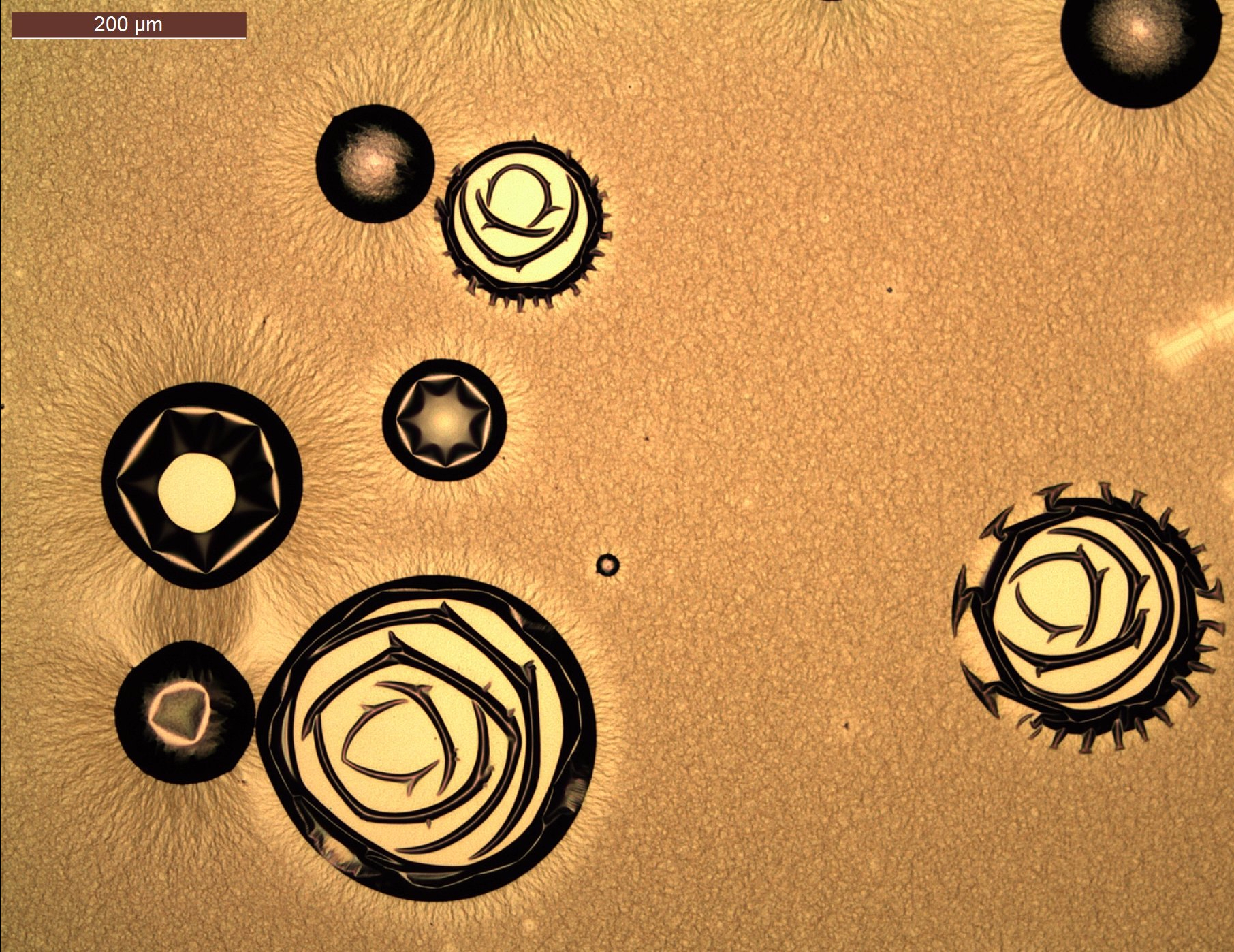
Micrometer-size gold roses – VLYETSTRA, Nynke
Top view of a not-so-perfect gold thin film. The aim was to deposit a 40-nm-thin film of titanium and gold by electron-beam evaporation, in order to pattern nanometer-size structures on the substrate. During the deposition on the glass substrate, which was covered with a polymer, the underlying polymer got attacked for an unclear reason and the growth of the film was disturbed. The result is a not at all smooth layer with many ‘very large’ gold clusters, which look like small microscale roses. Nice artwork as a result of a failed process. This image was taken with an optical microscope and a 10x objective.
Prize: Polaroid Snap shot & Professional PhotoShoot

- Dr. Iris Sancho presents a card game to introduce “Women in Science”
BEST IN-SCIENCE: Morphology of sensory neurons in vivo in Drosophila larvae.
Dendrite morphology of sensory neurons of 3rd instar larvae and epithelial cells by using a GFP enhancer trap visualizing an endogenous protein expression at 96h AEL (after egg laying). You clearly see an expression gradient from the central to the lateral areas of the receptive field with very little expression along the receptive field barrier indicating this epithelial secreted protein influences the growth of sensory neuron dendrites within their receptive field. Neurons were visualized by using a pan-sensory specific Gal4-driver and the expression of tdTomato. Image of 3rd instar larval sensory neurons was taken in vivo by confocal stack microscopy (Zeiss LSM700) and a 40x objective. Scale bar: 50 µm.
BEST OUT-SCIENCE: Looking into the Universe
The photo was taken in the Deutsches Museum while exploring the exhibition dedicated to the Universe and our place in it. This is one of the many boxes inside of which separate stars and constellations were represented. However, due to reflections and lights inside the box as well as outside of it, we can see the small model of our endless Universe. The photo was taken with Fujifilm X70 camera (18.5mm-F2.8 lens), outlines of which can be seen in the Space.
Prize: 2 Cinema tickets + 2 cocktails
#CERFA_Bayern feels grateful and happy about the warm reception of #Photon2017, and we hope that you enjoyed the experience as much as we did. We think that all of you are winners after having reached this phase, and that is why we would like to dedicate this space to all the exhibited ones and reserves (R).
Enjoy the trip around Art&Science!
THANKS!
Scroll down to reveal their respective captions. They will help you understand the images!
- Dr. Natalia Balcázar, Talking about Gender Perspective
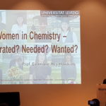
- Prof. Evamarie Hey-Hawkings, University of Leipzig talks about Women in Chemistry and her personal experience as successful scientist
- Prof. Evamarie Hey-Hawkings, University of Leipzig talks about Women in Chemistry and her personal experience as successful scientist
- Prof. Daniela Gordfarb, Institute Weizmann, Israel, talks about her current topic of research
- Prof. Daniela Gordfarb, Institute Weizmann, Israel, talks about her current topic of research
- Dr. Iris Sancho presents a card game to introduce “Women in Science”
- Dr. Agnes Benardeau, Scientist Manager, Bayer AG – Role models of succcess in industry
- Dr. Agnes Benardeau, Scientist Manager, Bayer AG
- Dr. Sarah Theisen, Product Safety Specialist Global HSE, Siegwerk AG – ole models of succcess in industry
- Dr. Sarah Theisen, Product Safety Specialist Global HSE, Siegwerk AG – ole models of succcess in industry
- Dr. Sarah Theisen, Product Safety Specialist Global HSE, Siegwerk AG – ole models of succcess in industry
- Discussion panel: How can german Mentoring Programs help for Career Planning – with Mw. Susanne Müller, Ms. Rosemarie Fleck, Ms. Ekaterina Masetkina
CAPTIONS:
01 Captain America’s shield made of light
Duo-chromatic laser beam composed of 515nm (green) and 670nm (red) wavelengths as it exits a multimode optical fiber. Such an illumination technic allows to combine two imaging approaches in space and time. Red and green beams have different size and shape due to wavelength-dependent dispersion. ‘Noisy’ structure of light is formed by random interaction of different modes in multimode fiber.
02 Reverse time by adding color in your life
Top of view of lung cut from aging mice (12 mouths): Cellular aging is marked by Senescence associated beta-galactosidase staining. The cells in the process of aging will be marked blue in the cytoplasm. The nuclei are colored in purple with hematoxylin-staining, to count the number of cells. When I started my analysis, I found it very pretty, difference with presence of blue that correspond to the aging, with the passage of time. And this rainbow effect, which is an artefact or experimental error. But it gives back the mouse, and recalls expression “After the rain, there is always a rainbow”. It made me smile despite an irreversible mechanism called aging, and the rainbow that gives unreal impression. I find that this image despite that it represents the aging, gives an imaginary dimension. Image taken with MIRAX SCAN, Carl Zeiss microscope with a 20X objective.
03 The Heart of a Hummingbird
Human cancer cell line, expressing an introduced mutant fms-related tyrosine kinase 3 (FLT3) receptor. The mutant receptor was detected in an acute myeloid leukemia (AML) patient. Although inducing constitutive downstream signaling, the mutant FLT3 receptor (labelled with an anti-FLT3 antibody, blue) was located similar to the wild type form – dominantly at the cell membrane and perinuclear. Glycoconjugates were targeted with a wheat germ agglutinin (WGA) antibody (turquoise). DNA (white) was labelled with 4’,6-Diamidin-2-phenylindol (DAPI). Image was taken using a confocal laser scanning microscope. It reminds me of a capture of a hummingbird that hovers next to a flower – the wings full of power and its heart full of energy.
05 Bubble bobble
Mitochondria, the cellular energy production plants in all eukaryotic cells, are related to multiple human diseases. The picture shows the mitochondrial outer membrane protein TOMM20 (red) and the component of Complex IV COXI (red) in the inner mitochondrial membrane. Typical light microscopes cannot distinguish between different mitochondrial compartments and its analysis was restricted to biochemical studies. The resolution of the microscope allows us to distinguish individual subunits of the OXPHOS system and view them as they are organized inside mitochondria. Single mitochondria resemble soap bubbles that took me to my childhood with the famous 80s’ videogame Bubble bobble. Image taken with Leica Confocal gSTED with 60x objective and digital magnification of the area.
06 Now You See Me
G-protein coupled receptors (GPCRs) are life’s favorite way to receive and transmit information across cellular membranes. A stimulus arrives and these proteins quickly rearrange their structure. Stimuli can range from smells, tastes, hormones and neurotransmitters to the most important one for this (or any) photo: light! Yes, right now, the GPCRs in your retina are rearranging to let your brain know that photons are arriving. MD simulations and kinetic analysis help us visualize 1) how much the active (green) and inactive (red) states differ and 2) the conformational flexibility within each ensemble as predicted by thermodynamics. Image rendered on VMD with 2×400 Boltzmann-weighted, cropped-overlays of the active and inactive ensembles.
07 Multimodal microscopy of a mouse ear
Hybrid label-free examination of an in vivo mouse ear by combining optical-resolution optoacoustic (photoacoustic) with non-linear optical microscopy modalities. Microvasculature is revealed by optoacoustic microscopy (red), bulb regions of hair follicles by two-photon excitation auto-fluorescence (white), collagen in the underlying dermis layer by second harmonic generation (green), and tissue morphology as well as keratinocytes by third harmonic generation microscopy (blue). Field of view is 3.33 x 2.47 mm². Microscopy images were acquired using a custom-built multimodal microscope enabling an insight into the beautiful complexity of biological tissue at the microscale.
09 Sensing the Dance
Cranial sensory nerve cells expressing the green fluorescence protein (GFP) and Cy3 labelled antibody against the p75 receptor due to a virus co-injected into whiskers and transported internally to the midbrain of the rat. These combined images could be dancers sensing ‘the music groove’ – claimed to be an understanding of rhythmic patterning that stimulates dancing on the part of the listener. Image taken with a confocal microscope (LSM 710) using a plan-Apo 40 9 NA 0.95 objective (Carl Zeiss, Germany).
11 A Ladder to the brain
Ventral nerve cord of a 96h old Drosophila larva expressing tdTomato in Class 4 Sensory neurons showing ladder-like axon projections and their downstream partners expressing GFP. One can perfectly see the overlap of the two neuron types. Every “step” of the ladder is responsible for one segment of the larva and transduces noxious stimuli, such as pain and heat, into the larval brain. Drosophila larvae react with a stereotype nocifensive escape behavior to those stimuli, by rolling and bending, trying to escape stings from parasitic wasps in nature. All animals need sensory systems to trigger adequate responses and ensure survival. Image taken with a Confocal Laser Microscope and a 40x oil objective.
12 In the Crowd
This takes place in a brain, a fish brain. The pink one is a cell and could be any other cell, makes itself comfortable throughout the crowd. There, every cell has a blue nucleus. Who knows what they are all telling to each other? Image taken with a confocal microscope with a 63x objective.
13 “Über den Tellerrand blicken”
Literally the German title “Über den Tellerrand blicken” means “to look over the edge of the plate” and the figurative meaning is to think outside of the box and extend one’s horizon. The challenges of a PhD often force me to “look over the edge of the plate” which consequently result – in the best case – in evolvement of my skills, new insights and development of my personality. I took the photo during our Halloween party in October 2016. I myself created the brain out of a watermelon and for the purpose of Halloween it was resting on a plate to scare my friends a little bit. The blue plastic eye balls and the bony hand were the perfect features to be successful with this task.
14 Living art – Genetically engineered bacteria as paint
This photo shows a set of our 5 illustrations drawn in the lab depicting Charles Darwin, Albert Einstein and two Mandalas. What at first sight appears to be some drawing done with a black marker pen, is actually living, genetically engineered bacteria that produce the deep purple pigment “Violacein”1. Instead of “Oil on canvas”, we use our technique “Bacteria on jelly”. In order to achieve that, the genes responsible for generating Violacein were transferred from the bacterium Chromobacerium violaceum to a common laboratory strain of Escherichia coli, providing it with the ability to produce vast amounts of Violacein. Accordingly, a highly-diluted, colorless bacterial suspension of our strain was applied with ordinary brushes to the “canvas”, which in our case is a 15 cm-diameter petri dish, containing a jelly-like substrate that the bacteria love to grow on. After 24 hours of incubation at 37°C, bacteria had overgrown, and the areas that were marked with the brushes have produced a large amount of the beautiful purple pigment, revealing the motif that was drawn by us. Interestingly, Violacein has several other potential applications beyond using it as paint: Since its optical spectrum extends into the near-infrared range, our strain of bacteria can easily be detected with a new imaging modality called “Optoacoustics”, in which absorbing molecules are being illuminated with short laser pulses creating heat and thereby ultrasonic pressures waves, that can be reconstructed into images. Furthermore, we have shown that the molecule itself has cytotoxic activity against certain cancer cell lines. In the future, our engineered bacteria could potentially serve as theranostic vehicles circulating in the body, enriching in tumors and thereby enabling their imaging via optoacoustic and at the same time killing off tumor cells. doi:10.1038/srep11048 (2015).
15 A day in the life of a model organism
Spontaneous snapshot of Arabidopsis thaliana seedlings growing on MS (Murashige and Skoog) agar-plate. Plant botanists began to study the thale cress Arabidopsis thaliana in the early 1900s, and nowadays is widely used as a model organism in plant biology, though, is not of major agronomical significance it belongs to an agronomical important family- Brassicaceae, and it offers important advantages for basic research, among them; its small genome, its life cycle can be completed in 6 weeks, and as illustrated in the picture, easily grows in a restricted space. Image taken with a Canon 550d.
16 To be or not to be… a neuron
This picture shows two cell types derived from neural stem cells isolated from the brain of a mouse embryo at 13.5 days of development. When cultured in an appropriate culture medium for enough days, neural stem cells can develop into neurons (positive for beta tubulin III, in green) or astrocytes (positive for the glial fibrillary acidic protein, in red). Neurons are able to establish complex synaptic circuits that allow the processing and transmission of information. Astrocytes are especially important because they support neurons and provide them with nutrients. Here, a group of astrocytes are embracing a neuron, maybe helping this young cell to mature and start making synaptic contacts with other neurons. Image taken with a confocal microscope at a 40x magnification.
17 Green Fat
This a fluorescent image of differentiated adipocytes expressing GFP. The cells were electroporated with a GFP- expression plasmid at the preadipoczytes stage and differentiated using a cocktail of different chemicals and proteins. Luckily, the electroporation procedure did not stress the cells and they differentiated normally. This is usually not the case with other transfection methods. This picture was a part of electroporation optimization experiment. The bright green dots are lipid droplets containing GFP and resemble a bit like tiny green stars trapped in a web of cells. Image taken from a standard inverted cell culture microscope with fluorescent light source at 4x.
18 Science meets Art. A Bavarian – Spanish –co-production
Scientific images very often disclose fantastic motifs. Looking at them open mindedly without knowing the background of the image it can stimulate the viewers’ imagination. That happens when we take the micrographs of a thin film of antipyretic drug Indomethacin®. This material is of physical interest because it forms a disordered “glassy state” material like silica glass known from windows which usually do not crystalize. They are permanent liquids but flow so slowly that they seem to be solid. Their molecules can rotate and translate almost randomly around the equilibrium position. But what happens, when the system is cooled down and the movements of the molecules are frozen? During transportation by air plane from our Spanish collaborator, the samples have been exposed to extreme conditions (pressure, temperature, humidity) which evoke abnormal crystallization. Fortunately this “accident” led to an amazing result: Disordered systems can crystalize in a high ordered system. Even if something went wrong in science it can be worthwhile… Images have been taken with an optical Microscopy Leica DM 6000 in polarization mode without any follow-up treatment.
19 Fly Brain on Fire?
High-magnification of a fruit fly’s brain focusing on the lamina, a region that processes visual information coming directly from the fly’s eye. The lamina contains an exquisitely arranged array of ~750 units known as cartridges. This image shows 8 cartridges in red (after immunolabeling of a synaptic protein) and a few neurons in yellow (after expression of a fluorescent protein). The axons within the cartridges (bottom) glow like they are on fire and the synapses (in red) shine like sparks. The burning is, however, an illusion since the balloon-like somata (top) do not seem to fly apart. Image was taken with a Confocal Microscope Leica TCS SP8, 63x Objective, zoom=4.5. A neuron’s soma diameter is ~3 micrometers.
20 Early developmental moments
Following germination, shoots of most plants pass through a phase of vegetative growth, during which plants form leaves and rapidly increase photosynthetic capacity and their size and mass. After the vegetative phase is completed, plants switch to reproductive growth, where the plant acquires reproductive competence. In the picture it can be observed the juvenile phase of vegetative growth of the model plant Arabidopsis thaliana. Image taken with a Canon 550d camera using an inverse ring.
RESERVES:
R1 One and only
View of an Intracytoplasmic Sperm Injection (ICSI), in which a single sperm cell is deliberately injected into an in vitro matured bovine oocyte bypassing all natural barriers. Injection pipette goes through Zona Pelucida breaking the cell membrane. Ooplasm is first aspirated and then softly placed back carrying the choosed sperm. Hours later sperm decondenses its DNA and 2 pronucleous are fused into a zygote. This is the begining of a new life. This reminds me of Aristotle: Three is the most perfect number,—it is the first of numbers, for of one we do not speak as a number, of two we say both, but three is the first number of which we say all. Moreover, it has a beginning, a middle, and an end. Image taken with a Nikon Eclipse inverted microscope with a 200x Objective.
R2 Robot, please make us happy!
Robots will be part of our society and will live with us to help us in our daily life and entertainment us as well. These robots will learn to play football even better than humans; and robotic Worldcups will collapse press media and internet. Picture is taken during a robocup competition at the Institute of Cognitive Systems, Technische Universität München. The robot in the image is called NAO. It is the official robot for the Robocup. These robots autonomously play football using fast artificial intelligent algorithms while dozens of followers emotionally shout to support their teams. The camera employed is a Canon EOS 500D Digital camera with a Canon EF-S 18-55mm IS lens and edited with Photoshop CS6 v13.0.
R3 A camaleon in a liposome zoo
Cross-secion of liposomes labeled with DSPE-Rhodamine Red encapsulating carboxyfluorescein (GFP fluorescent properties) observed by confocal laser scanning microscopy. Liposomes were generated by overnight swelling of a phospholipid cake with an aqueous solution containing carboxifluorescein and sucrose. Liposomes were sedimented on top of a microscope coverslip via a sucrose-glucose gradient. This image reminds us how infenitely diverse life is. Liposomes are a synthetic version of a typical cell plasma membrane, the basic containers that allow life to take place. As in the living world, liposomes originating from a single homogeneous mixture of lipids, show a great heterogenous spectrum of shapes, sizes and content. The camaleon in the right bottom side of this picture is just one more member inside a beautiful ocean of diversity.
R4 Sakura
By the end of March, the parks in Japan become pink with the petals of the Cherry Blossom and get crowded with people celebrating the Cherry Blossom season under the trees with friends or family. In Japanese culture, the cherry blossom represents the fragility and beauty of life and it is present everywhere as a reminder that life is as beauty as ephemeral. Although this image might remind to a cherry blossom, it is actually a neuromuscular junction from a fruit fly. Every pink dot is a contact between the neuron and the muscle and all together, when property active, will make the fly move. The same way our neuromuscular junctions will make us run, jump, dance under the cherry trees. Image taken with a confocal microscope, with 40x oil immersion objective.
R5 Sand stars
This picture was taken close to the science city complex on top of the Haleakala volcano in Maui, Hawaii. The Advanced Electro-Optical System in the observatory in about 3050 m altitude provides excellent astronomical seeing conditions. However, you can also look down and discover the most amazing stars under your own feet.
R6 Life in a Bubble
Picture of air bubbles in a slide which has RNA in-situ hybridization (method to find expression of a gene) samples of Drosophila (fruit fly) embryos. While searching for embryos in the slide these small air bubbles caught my interest. The scientific samples and the air bubbles are like present scientific community and the common people. While trying to answer the big scientific questions, scientists are failing to communicate with the general public. This lead to two bubbles, scientific bubble and general public bubble. I wish these bubbles just fuse together and form a bubble. But I fear that the distance between the bubbles are increasing due to the current political situation on this earth. I believe that by breaking these bubbles we can avoid events like the protest against vaccination. Image taken with a stereomicroscope and the mounting medium is glycerol.
R7 Generation-R
Generation R is the first generation of Robotic Natives. Nowadays we have kids that interact even without thinking with tablets and touch screens. We will only have to wait for our grandchildren to observe a shift in the society behaviour regarding human like robots. The generation R will naturally interact with robotic systems such as smart homes, assistive humanoids, robotic bartenders or administrative and legal robots. Hold on!, maybe we are already experiencing the robotic era. This image captures an spontaneous encounter between a 6 year old kid and iCub during the open day at the Institute of Cognitive System (ICS), Technical University of Munich. The iCub robot proportions are as a 3.5 years old and was built as a result of a European project consortium. Camera: Canon EOS 500D with EF-S 18-55mm IS lens. Blur and colour effects have been added using Photoshop CS6.
