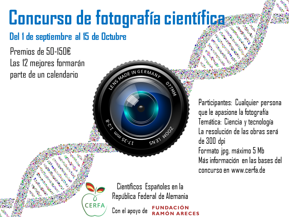
CERFA lanzó su segundo concurso de fotografía científica en septiembre y octubre de 2022 para aquellos investigadores y no investigadores interesados en difundir diferentes enfoques sobre la ciencia y la tecnología a partir de la fotografía. Con las fotografías participantes, hemos confeccionado un calendario de mesa para 2023 que puedes descargar aquí (tamaño A6 con margen suficiente para recortar y poner anillas en la parte superior).
Las bases completas del concurso se encuentran aquí.
Los/as ganadores/as anunciados al final de la Asamblea General de CERFA el pasado 13 de noviembre fueron los siguientes:
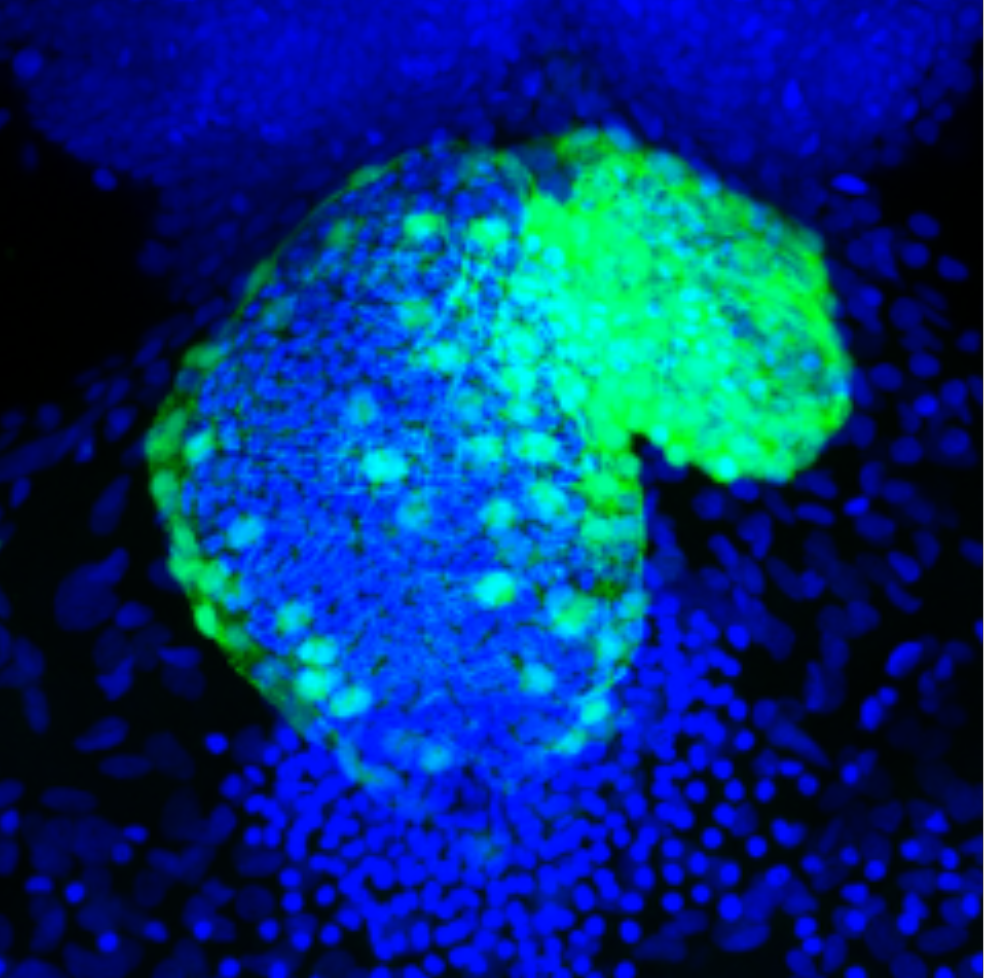
Bombeando vida – Pumping life (1er premio del jurado)
Sergio G. San Miguel
Universidad de Potsdam, Postdam, Alemania/Germany.
Afiliación actual: Max Planck Institute for Infection Biology, Berlin, Alemania/Germany.
Descripción:
En este proyecto, estábamos interesados en entender cómo se forma el tejido cardiaco en las primeras horas de vida del Pez Cebra. El Pez Cebra es un excelente modelo para estudiar la formación del corazón. Los embriones, al ser tan pequeños, pueden toleras mutaciones o drogas que afectan específicamente al corazón, ya que el oxígeno y nutrientes se difunden sin necesidad del sistema cardiaco. La foto está tomada con microscopio confocal de un corazón de Pez Cebra (Danio rerio) a las 48 horas después de eclosionar. Células del miocardio están marcadas con proteína fluorescente verde (GFP). Núcleo de todas las células está marcado con la molécula fluorescente 4 ‘,6-diamidino-2-fenilindol (DAPI). Este representa un corazón del grupo control.
Description:
In this project, we were interested in understanding how heart tissue is formed in the first hours of life of the zebrafish. The zebrafish is an excellent model to study heart formation. The embryos, being so small, can tolerate mutations or drugs that specifically affect the heart, since oxygen and nutrients are diffused without the need of the cardiac system. The photo is taken with confocal microscopy of a zebrafish (Danio rerio) heart 48 hours after hatching. Myocardial cells are labeled with green fluorescent protein (GFP). Nuclei of all cells are labeled with the fluorescent molecule 4′,6-diamidino-2-phenylindole (DAPI). This represents a heart of the control group.
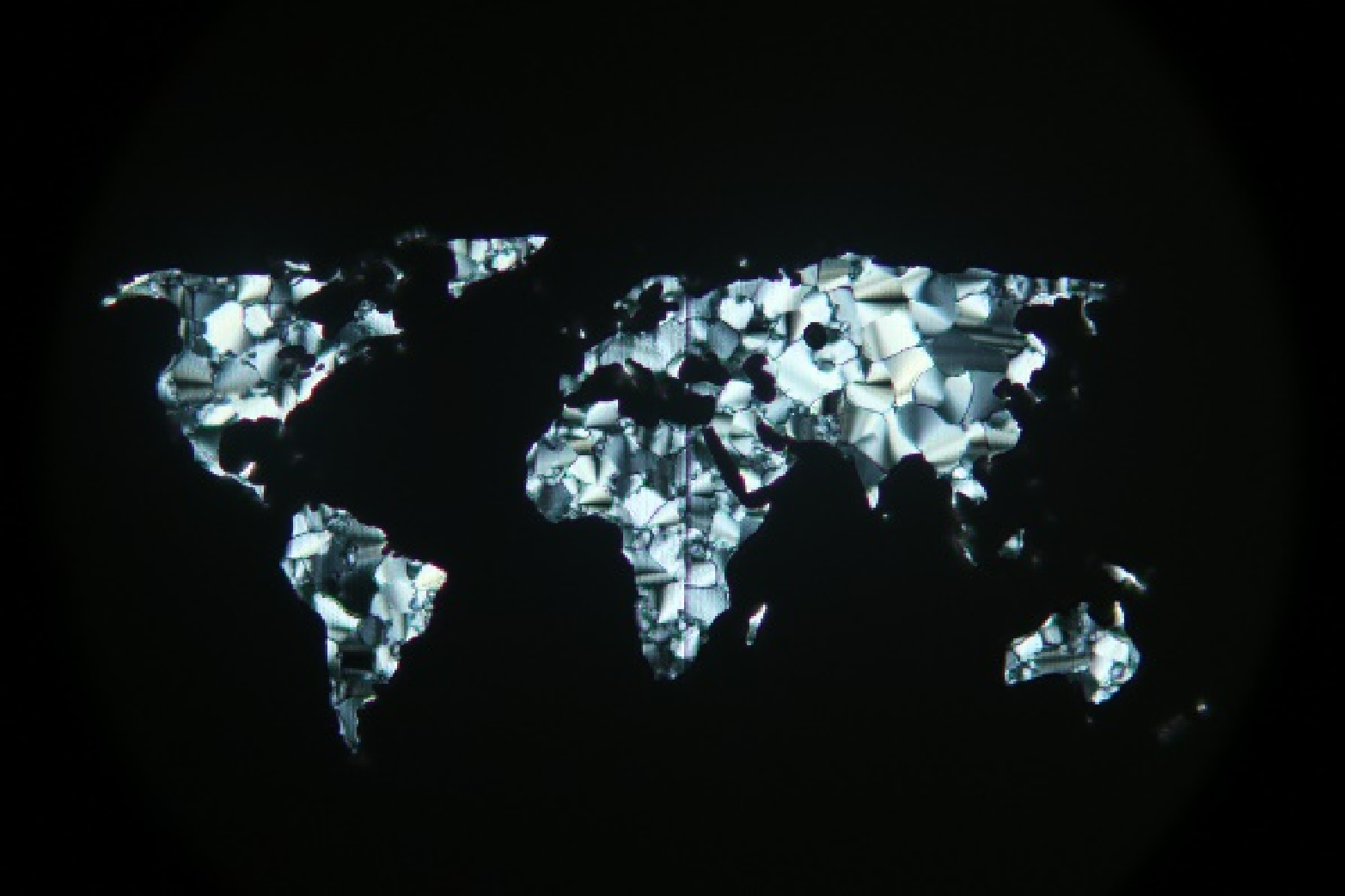
Mapamundi cristal líquido – Liquid crystalline micropatterned world map (2º premio del jurado y premio del público)
Eva Blasco y Joel Monti
Heidelberg University, Heidelberg, Alemania/Germany.
Descripción:
Demonstración de la microimpresión láser utilizando un material cristal líquido para la fabricación de patrones altamente definidos.
Description:
We demonstrate the succesful laser microprinting using a liquid crystalline material for the fabrication of highly defined patterns.

Biomímesis: el camuflaje del camaleón – Biomimicry: the camouflage of the chameleon (Premio del público)
Rubén Rodríguez Puertas
Universidad de Almería, Almería, España/Spain.
Descripción:
La fotografía muestra una de las principales características del camaleón, su capacidad de controlar la pigmentación de su piel, y por tanto, de camuflarse. Este proceso está siendo analizado por la ciencia (principalmente la ingeniería) y aplicado a distintos objetos de nuestra vida diaria (viviendas, murallas, cortinas, vidrios…) que, a través de las llamadas pieles eléctricas, pueden ahorrar energía y cambiar de color ajustándose al entorno.
Description:
The photograph shows one of the main characteristics of the chameleon, its ability to control the pigmentation of its skin, and therefore, to camouflage itself. This process is being analyzed by science (mainly engineering) and applied to different objects in our daily life (homes, walls, curtains, glass…) which, through so-called electric skins, can save energy and change color by adjusting to the environment.
Agradecemos la participación de todos los concursantes y os dejamos a continuación el resto de fotografías:

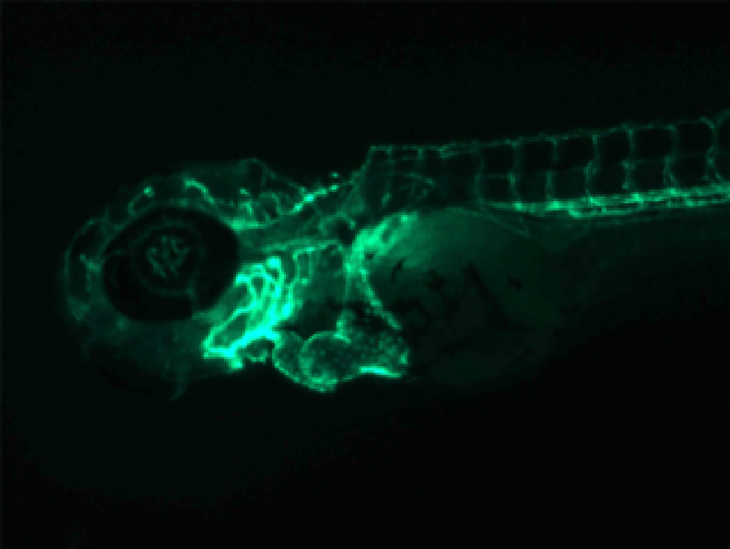
Embriones de pez cebra – Zebrafish embryos
Amaia Jauregi Miguel
Linköping University, Linköping, Suecia/Sweden.
Descripción:
El pez cebra es un miembro de la familia de los peces carpa. Este organismo es usado como modelo para estudiar el desarrollo de los vertebrados debido a que el embrión es transparente y se desarrolla rápidamente fuera de su madre. En la foto, se pueden observar dos embriones de pez cebra en el momento de la eclosión, al tercer día post-fecundación.
Description:
Zebrafish are members of the carp fish family. Embryos of this organism are transparent and develop rapidly outside their mothers, making them an excellent model for studying vertebrate development. Two embryonic zebrafish can be seen in the photo as they hatch on the third day after fertilization.

Bïrgerpark, Bremen
Daniel García Rodríguez
Evotec, Hamburg, Alemania/Germany
Descripción:
El Prof. Dr. Simon Eickhoff (Institut für Neurowissenschaften und Medizin INM) estudió los efectos a nivel neuronal que tenían dos imágenes bastante contrarias: una playa tranquila y virgen, sin muchos elementos destacables en el paisaje; y por el contrario, una avenida repleta de objetos inmóviles y móviles (semáforos, edificios, personas, coches…). La primera imagen más tranquila, a través de las cortezas sensoriales visual y auditiva, era resultado de una activación posterior de áreas como la corteza prefrontal (ligada a emociones) y en último lugar a cambios celulares y moleculares como puede ser el aumento de serotonina y, por tanto, de bienestar y felicidad. Tanto la pareja de ancianitos como el mismo espectador (y servidor), reciben una dosis de este conocido neurotransmisor que, por unos minutos al menos, nos alegra el día. Fotografía analógica (Canon AE-1).
Description:
Prof. Dr. Simon Eickhoff (Institut für Neurowissenschaften und Medizin INM) studied the effects at the neuronal level of two quite opposite images: a quiet and virgin beach, without many notable elements in the landscape; and on the contrary, an avenue full of immobile and mobile objects (traffic lights, buildings, people, cars…). The calmer first image, through the visual and auditory sensory cortices, was the result of a subsequent activation of areas such as the prefrontal cortex (linked to emotions) and, lastly, of cellular and molecular changes such as the increase in serotonin and , therefore, of well-being and happiness. Both the elderly couple and the viewer (and server) receive a dose of this well-known neurotransmitter that, for a few minutes at least, makes our day. Analog photography (Canon AE-1).
Un esqueleto hecho de sangre – A skeleton made of blood
Sergio G. San Miguel
Universidad de Potsdam, Postdam, Alemania/Germany.
Afiliación actual: Max Planck Institute for Infection Biology, Berlin, Alemania/Germany.
Descripción:
En este proyecto, estábamos interesados en entender cómo se forma el tejido cardiaco en las primeras horas de vida del Pez Cebra. El Pez Cebra es un excelente modelo para estudiar la formación del corazón. Los embriones, al ser tan pequeños, pueden toleras mutaciones o drogas que afectan específicamente al corazón, ya que el oxígeno y nutrientes se difunden sin necesidad del sistema cardiaco. La foto está tomada con microscopio fluorescente invertido. En ella vemos la parte anterolateral de un Pez Cebra (Danio rerio) a las 48 horas después de eclosionar. Células del endocardio están marcadas con proteína fluorescente verde (GFP).
Description:
In this project, we were interested in understanding how heart tissue is formed in the first hours of life of the zebrafish. The zebrafish is an excellent model to study heart formation. The embryos, being so small, can tolerate mutations or drugs that specifically affect the heart, since oxygen and nutrients diffuse without the need of the cardiac system. The photo is taken with inverted fluorescent microscopy. It shows the anterolateral side of a zebrafish (Danio rerio) 48 hours after hatching. Endocardial cells are labeled with green fluorescent protein (GFP).

El inicio de la vida – The beginning of life
Rubén Rodríguez Puertas
Universidad de Almería, Almería, España/Spain.
Descripción:
La fotografía muestra el rol principal de las abejas: el de polinizadoras. Gracias a esta función le debemos gran parte de nuestras frutas y vegetales, y no solo nosotros, la especie humana, sino todos los seres vivos de su alrededor. Por ello, con este rol que cumplen en su aparentemente sencillo vuelo de flor en flor, crean vida. Por estos motivos es importante divulgar más conocimiento científico sobre este importante insecto.
Description:
The photograph shows the main role of bees: that of pollinators. Thanks to this function we owe a large part of our fruits and vegetables, and not only we, the human species, but all the living beings around them. For this reason, with this role that they fulfill in their apparently simple flight from flower to flower, they create life. For these reasons it is important to spread more scientific knowledge about this important insect.
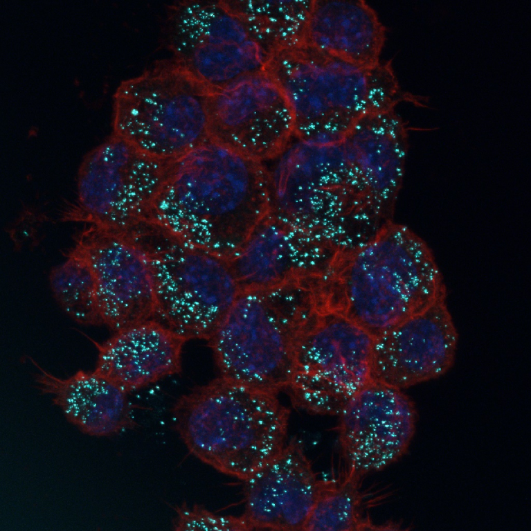
Macrófagos de ratón con nanopartículas de hierro – Murine macrophages with ferrous nanoparticles
María Teresa Marcos Almaraz
Institut Lavoisier Versailles, Versailles, Francia/France
Descripción:
Proyección máxima de microscopía confocal de la entrada en macrófagos de ratón de nanopartículas de hierro tipo nanoMOFs diseñadas para el encapsulamiento de fármacos, tras las primeras 24 horas de la adición al medio de cultivo de las nanopartículas, preparadas en solución fisiológica. En color brillante, se observan las nanopartículas, que autoflorecen debido a la excitación directa con el láser. En rojo, la membrana celular marcada con phalloidina y, en azul (debido al marcaje con DAPI), los núcleos celulares.
Description:
Maximum intensity projection of confocal microscopy picture showing the entrance of ferrous nanoparticles nanoMOF into murine macrophages. Nanoparticles were designed to encapsulate drugs and added to the culture. Picture was captured after 24h. Nanoparticles can be seen in a brilliant colour since they autofluoresce due to the direct illumination by the laser. In red, cell membranes are stained for phalloidin, and blue, cell nuclei are shown (DAPI staining).

Dagebüll, Schleswig-Holstein
Daniel García Rodríguez
Evotec, Hamburg, Alemania/Germany
Descripción:
El famoso mar del Norte, bonito y especial, pero a la vez, impredecible y aleatorio. En la foto se puede observar como un grupo de personas camina plácida y tranquilamente por una superficie que, hace literalmente cuestión de horas como mucho, era territorio custodiado por el mar.
El hecho de que este mar europeo sea tan distinto y peculiar con otros, es su posición estratégica, ya que se encuentra entre las Islas Británicas, la península de Escandinavia y el territorio europeo continental. Esto hace que por el sur (Canal de la Mancha) llegue una corriente y por el norte otra (espacio que queda entre Escocia y Noruega), ocasionando estas mareas tan extremas. Por suerte, en la mayoría de casos, gracias a estudios de mareas, se pueden obtener los denominados calendarios de mareas, gracias a los cuales puedes campar a tus anchas con más (o menos) tranquilidad. Fotografía digital (Nikon D3500).
Description:
The famous North Sea, beautiful and special, but at the same time, unpredictable and random. In the photo you can see how a group of people walk placidly and calmly on a surface that, literally a matter of hours ago, was territory guarded by the sea. The fact that this European sea is so different and peculiar with others, is its strategic position, since it is located between the British Isles, the Scandinavian peninsula and the continental European territory. This means that one current arrives from the south (English Channel) and another from the north (space between Scotland and Norway), causing these extreme tides. Luckily, in most cases, thanks to tide studies, so-called tide calendars can be obtained, thanks to which you can roam freely with more (or less) peace of mind. Digital photography (Nikon D3500).
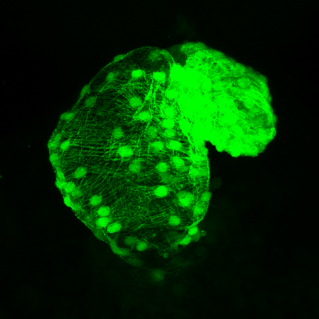
Luciérnagas cardiacas – Cardic fireflies
Sergio G. San Miguel
Universidad de Potsdam, Postdam, Alemania/Germany.
Afiliación actual: Max Planck Institute for Infection Biology, Berlin, Alemania/Germany.
Descripción:
En este proyecto, estábamos interesados en entender cómo se forma el tejido cardiaco en las primeras horas de vida del Pez Cebra. El Pez Cebra es un excelente modelo para estudiar la formación del corazón. Los embriones, al ser tan pequeños, pueden toleras mutaciones o drogas que afectan específicamente al corazón, ya que el oxígeno y nutrientes se difunden sin necesidad del sistema cardiaco. La foto está tomada con microscopio confocal de un corazón de Pez Cebra (Danio rerio) a las 48 horas después de eclosionar. Células del miocardio están marcadas con proteína fluorescente verde (GFP). Este representa un corazón del grupo control.
Description:
In this project, we were interested in understanding how heart tissue is formed in the first hours of life of the zebrafish. The zebrafish is an excellent model to study heart formation. The embryos, being so small, can tolerate mutations or drugs that specifically affect the heart, since oxygen and nutrients are diffused without the need of the cardiac system. The photo is taken with confocal microscopy of a zebrafish (Danio rerio) heart 48 hours after hatching. Myocardial cells are labeled with green fluorescent protein (GFP). This represents a heart from the control group.
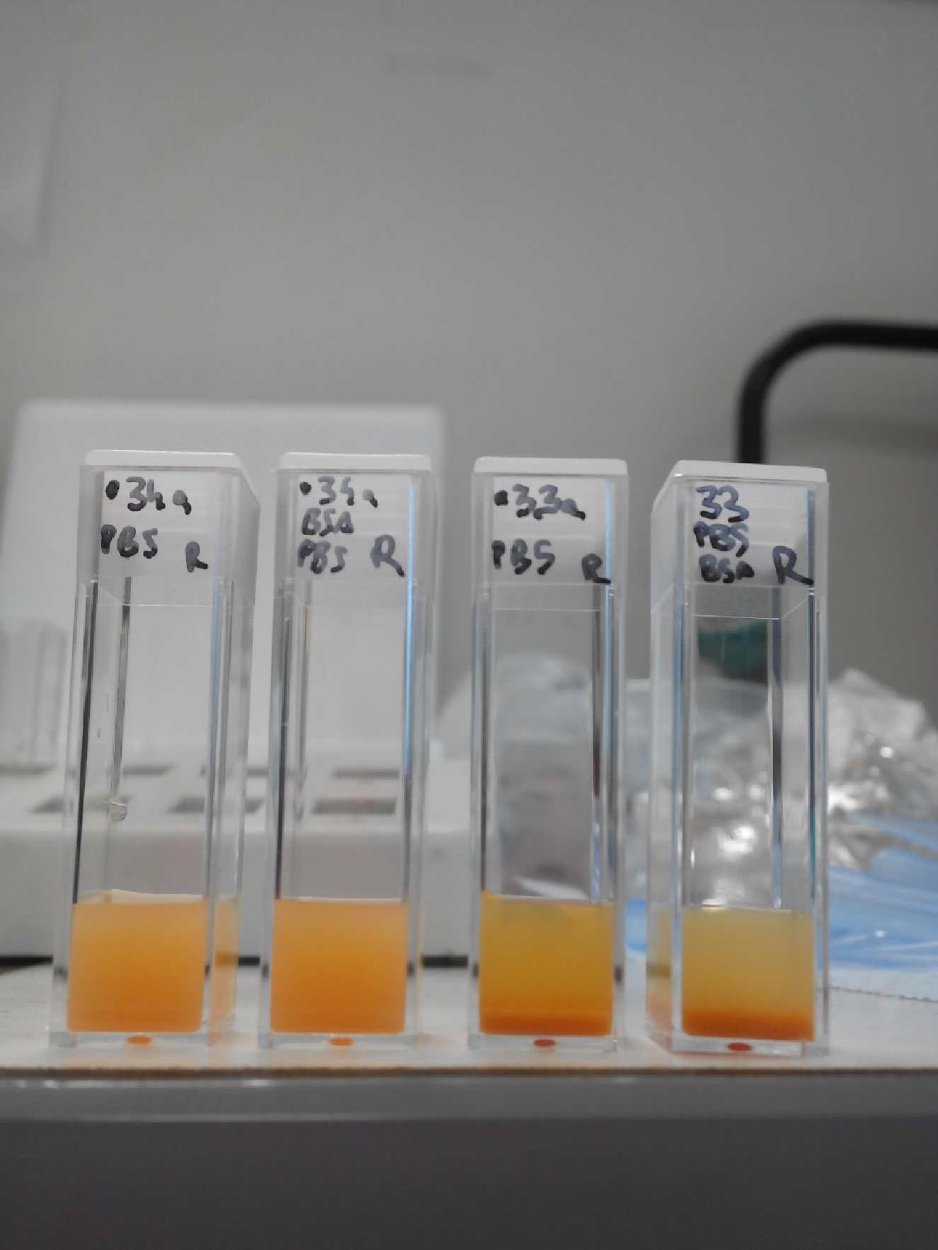
Nanopartículas de hierro – Ferrous nanoparticles
María Teresa Marcos Almaraz
Institut Lavoisier Versailles, Versailles, Francia/France
Descripción:
Comparativa de la estabilidad coloidal en dos soluciones fisiológicas (PBS y PBS+BSA) de dos tipos de nanopartículas de hierro (nanoMOF núm. 33 y nanoMOF núm. 34) diseñadas para el encapsulamiento de fármacos.
Description:
Comparison of the coloidal stability of two ferrous nanoparticles (nanoMOF No.33 and nanoMOF No.34) in two physiological solutions (PBS and PBS+BSA). Nanoparticles were designed to encapsulate drugs.
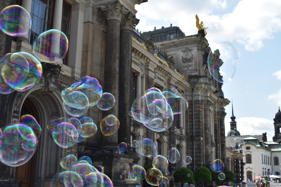
Terrassenufer en Dresden, Sajonia
Daniel García Rodríguez
Evotec, Hamburg, Alemania/Germany
Descripción:
Desde siempre, hacer pompas de jabón ha sido el entretenimiento principal y que más tiempo nos ha alegrado en nuestra infancia. ¿Pero cómo se hace posible que existan? Pura Física. Las pompas de jabón existen gracias al llamado efecto Gibbs-Marangoni y la propiedad de tensión superficial. Para explicar esta última, hay que imaginar las moléculas de agua en un vaso, de las cuales las que se encuentran más arriba en contacto con el aire, forman una interfaz agua-aire que crea dicha tensión superficial. Para crear burbujas, es necesario disminuir esa tensión, lo cual se consigue gracias a moléculas como el jabón (cabeza hidrófila que atrae esas moléculas de agua y una cola hidrofóbica que las repele). A estas moléculas se les denomina tensoactivas.
Y la pregunta del millón, ¿por qué pompas esféricas y no cuadradas? Simplemente la naturaleza hace la “ley del mínimo esfuerzo”, como se diría vulgarmente. La forma que encierra el mayor volumen en la menor superficie, la esfera. Fotografía digital (Nikon D3500).
Description:
Blowing bubbles has always been the main entertainment and the one that has made us happy the longest in our childhood. But how is it possible for them to exist? Pure Physics. Soap bubbles exist thanks to the so-called Gibbs-Marangoni effect and the property of surface tension. To explain the latter, imagine the water molecules in a glass, of which those higher up in contact with the air form a water-air interface that creates said surface tension. To create bubbles, it is necessary to reduce this tension, which is achieved thanks to molecules such as soap (a hydrophilic head that attracts these water molecules and a hydrophobic tail that repels them). These molecules are called surfactants. And the million dollar question, why spherical and not square bubbles? Simply nature makes the “law of minimum effort”, as it would be said vulgarly. The shape that encloses the largest volume in the smallest surface, the sphere. Digital photography (Nikon D3500).
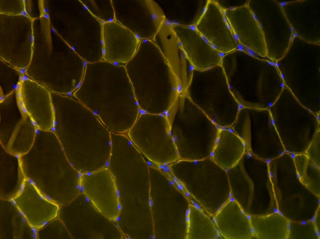
Músculo esquelético de corazón de ratón – Skeletal muscle of murine heart
María Teresa Marcos Almaraz
Institut de Myologie, Paris, Francia/France
Descripción:
Microscopía óptica a un aumento 20X de una sección de criostato de músculo esquelético de corazón de ratón. Los núcleos celulares están teñidos con DAPI y, en amarillo, se observa la colocalización de dos anticuerpos diferentes dirigidos contra el extremo N-terminal de la distrofina (Alexa 488) y contra la distrobrevina (Alexa Fluor 568).
Description:
Optical microsocpy with 20x augmentation of a section from the cryostat showing skeletal muscle from murine heart. Cell nuclei were stained with DAPI (blue) and the colocalization of two antibodies against the N-terminal of distrofin (Alexa 488) and distrobrevine (Alexa 568) are shown in yellow.